

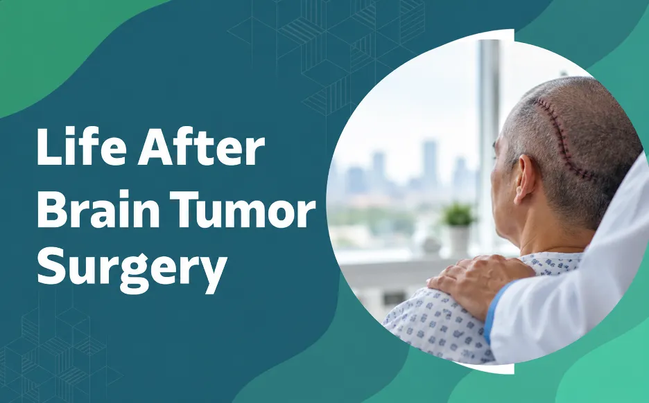
Life after brain tumor surgery follows a gradual path. The first two weeks bring fatigue, headaches, and swelling. Weeks two to six are about rebuilding energy and focus. By three months, many patients return to light work, and steady gains continue through the first year. Recovery depends on tumour type, location, and your overall health. Rehab, rest, good nutrition, and honest follow-ups improve outcomes. Report seizures, new weakness, or a sudden severe headache straight away.
Life after brain tumor surgery follows a gradual path. The first two weeks bring fatigue, headaches, and swelling. Weeks two to six are about rebuilding energy and focus. By three months, many patients return to light work, and steady gains continue through the first year. Recovery depends on tumour type, location, and your overall health. Rehab, rest, good nutrition, and honest follow-ups improve outcomes. Report seizures, new weakness, or a sudden severe headache straight away.
Life after brain tumor surgery rarely snaps back to normal overnight. The brain needs time to settle as swelling reduces and healthy tissue recovers around the surgical site. Early days are mostly about rest. Over the following weeks and months, energy returns, thinking sharpens, and everyday routines start to feel familiar again. Some patients bounce back quickly, while others need therapy and patience along the way.
According to Dr. Gurneet Singh Sawhney, Neurosurgeon in Mumbai,
“Recovery is a timeline, not a deadline. Two patients with the same surgery can heal at very different speeds, and both can be completely on track. What I ask patients to watch for is a sudden change, not the slow, uneven climb back.”
Recovering From Brain Tumor Surgery and Unsure What’s Normal? Get a clear, personalised recovery roadmap built around your case.
What to Expect in the First Two Weeks
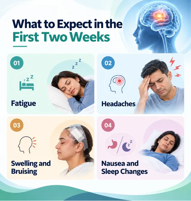
The first phase is the body’s repair window, and most of what you feel here is temporary. Knowing this in advance removes much of the fear that comes with the first days at home.
Fatigue
Deep tiredness is the most common early effect. The brain is using a large share of your energy to heal, so frequent rest is not laziness. It is part of the treatment.
Headaches
Mild to moderate headaches near the incision are expected and usually ease with prescribed medication and rest.
Swelling and Bruising
Scalp or facial swelling often peaks in the first few days, then settles as inflammation drops.
Nausea and Sleep Changes
Nausea linked to anaesthesia fades quickly. Broken sleep and vivid dreams are common and usually settle within a month.
Most patients are discharged within a few days to a week, depending on the procedure. Planning before and after brain tumor surgery shapes how gentle this phase feels, particularly when minimally invasive techniques are used.
Weeks 2 to 6: Rebuilding Energy and Routine
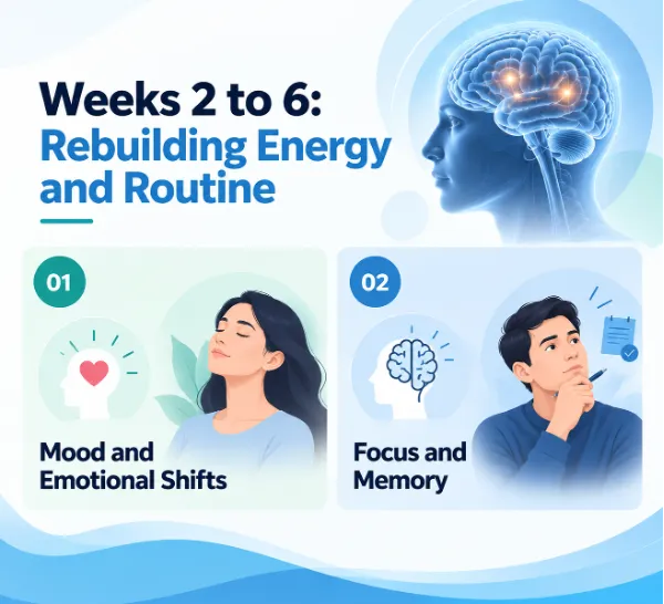
Energy stays low in this stretch, but small gains add up. Short walks, regular meals, and a steady sleep schedule do more for recovery than pushing too hard.
Mood and Emotional Shifts
Irritability, low mood, anxiety about scans, and unexpected tears are common. They reflect both healing tissue and the weight of a major diagnosis. Talking openly with family and your care team helps.
Focus and Memory
Concentrating for long periods feels harder, and short-term memory lapses are common. Working in short, rested bursts and using notes or reminders helps.
Weeks 6 to 12: Targeted Therapy Phase
This is when rehabilitation does its most important work. Depending on where the tumour was, you may need physical, speech, or cognitive therapy. Mild weakness, balance trouble, or word-finding difficulty often improves steadily with consistent practice.
For tumours near speech or movement areas, techniques such as awake craniotomy help protect those functions in real time during surgery, which often leads to smoother recovery afterwards.
Struggling With Memory, Speech, or Balance After Surgery? Early, targeted rehab can make a real difference.
Recovery Timeline: What Happens When
Healing follows a broad pattern, though pace varies with age, general health, tumour type, and location. Treat this table as a map, not a fixed schedule.
Timeframe | What Can Happen | What It Usually Means |
0–2 weeks | Fatigue, headaches, swelling, nausea | Normal early healing, managed at home |
2–6 weeks | Mood swings, poor focus, low stamina | Brain settling, gradual improvement expected |
6 weeks–3 months | Memory or speech gaps, mild weakness | Often responds well to targeted therapy |
3–12 months | Steady gains with occasional plateaus | Long-term recovery, monitored via scans |
Plateaus are not failures. Progress often arrives in bursts, with quiet stretches in between. Consistent rehab through those quieter phases protects the final result.
Life Beyond the First Year
For many patients, the first year is the steepest part of the climb. After that, recovery continues but more slowly, and the focus shifts to long-term wellbeing.
Follow-Up Scans
Regular MRI scans confirm healing and watch for recurrence. The schedule depends on tumour type and grade, so follow your surgeon’s plan closely, even when you feel well.
Return to Work and Daily Life
Many patients resume desk or light work within one to three months. Physically demanding jobs, driving, and sports take longer and need your surgeon’s approval. Some tumour types also need further treatment such as radiotherapy or medication, which can affect energy for a while.
Seizure Risk
Some patients, especially those with tumours near certain brain regions, may have seizures before or after surgery. Where seizures persist, expert review and options like epilepsy surgery can guide the safest next step.
How to Support Your Recovery
Small, consistent habits protect your result far more than most patients expect.
- Rest well: Sleep in a quiet, dim room and nap when needed.
- Eat for healing: Prioritise protein, fruit, vegetables, and plenty of fluids.
- Move gently: Short daily walks rebuild stamina and lift mood.
- Stay on schedule: Take medication on time and never skip follow-ups.
- Avoid strain: No heavy lifting, bending, or driving until cleared.
- Lean on others: Let family help with meals, transport, and reminders.
Want the Best Possible Outcome After Surgery? A structured follow-up plan with an experienced neurosurgeon keeps recovery on track.
When Should You See a Neurosurgeon After Brain Tumor Surgery?
Most patients heal steadily, but a few symptoms need urgent review. Catching a complication early makes it far easier to treat.
Get prompt specialist attention if you notice:
- A sudden, severe headache unlike your usual post-op ache
- A new seizure, twitching, or any loss of awareness
- Fresh weakness, numbness, or difficulty speaking
- Fever, redness, or fluid leaking from the incision
- Confusion, drowsiness, or vision that suddenly worsens
Guidance from the National Cancer Institute highlights that follow-up care and monitoring are central to long-term outcomes after brain tumour treatment. Reputable hospital resources such as the Mayo Clinic’s overview of craniotomy also explain what typical recovery looks like.
Dr. Gurneet Singh Sawhney, Neurosurgeon in Mumbai, stresses that honest follow-up drives long-term success. “The patients who recover best are the ones who tell me about small changes early. A symptom flagged at two weeks is far easier to manage than the same one ignored for two months.”
Frequently Asked Questions
How long does recovery after brain tumor surgery take?
Most patients improve noticeably over six to twelve weeks, though full recovery can take several months to a year depending on the tumour and surgery.
When can I return to work after brain tumor surgery?
Many patients resume light or desk work in four to twelve weeks. Physically demanding jobs and driving need your surgeon’s clearance.
Is fatigue normal after brain tumor surgery?
Yes. Deep tiredness is very common early on and usually eases gradually over several weeks.
Can brain tumor surgery affect memory or speech?
It can, especially when the tumour is near key areas, but therapy often restores much of the lost function over time.
When should I worry after brain tumor surgery?
Seek help immediately for new seizures, a sudden severe headache, fresh weakness, fever, or worsening confusion.
Reference:
- National Cancer Institute — Adult Brain Tumors Treatment (PDQ): https://www.cancer.gov/types/brain/patient/adult-brain-treatment-pdq
- Mayo Clinic — Craniotomy: https://www.mayoclinic.org/tests-procedures/craniotomy/about/pac-20394805
Disclaimer: The information shared in this content is for educational purposes only and not for promotional use.