

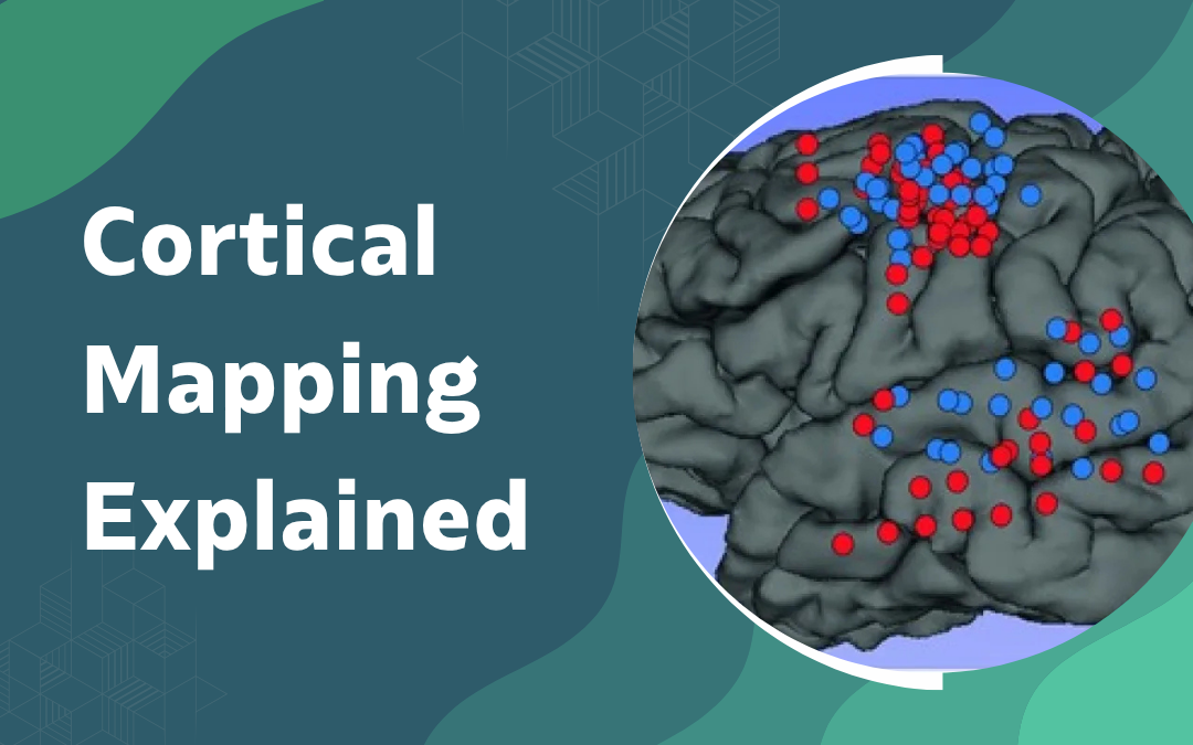
Cortical mapping identifies which brain areas control speech, movement, sensation and memory before tissue is removed during surgery. A small electrical current stimulates the brain’s surface while the patient performs simple tasks, and any disruption marks a region to avoid. This is why awake brain surgery exists; imaging alone can’t always predict where function sits once a tumour distorts normal anatomy. Mapping doesn’t remove risk, but it replaces guesswork with real-time feedback from the brain itself.
According to Dr. Gurneet Singh Sawhney, neurosurgeon in Mumbai, “Imaging tells me where a tumour sits, but only mapping tells me what the surrounding brain is actually doing, and that’s the difference that keeps a patient talking and walking normally afterwards.”
Protect Critical Brain Functions. Consult an Awake Brain Surgery Expert.
How Does Cortical Mapping Work During Surgery?
Mapping happens in real time once the skull is opened, using direct stimulation rather than imaging alone to confirm function.
- Direct Stimulation: a small probe delivers a brief electrical current to the exposed brain surface while the patient performs a task, and a pause in speech or a twitch in the hand marks that spot as functional tissue to avoid.
- Task Selection: tasks are chosen based on tumor location, so a lesion near the language center might involve naming pictures while one near the motor strip focuses on simple hand or foot movements instead.
- Sterile Grid Marking: numbered tags are placed directly on the brain surface at each tested point, building a live map that the surgical team references continuously as tissue removal proceeds.
- Continuous Feedback: an anesthesiologist and a neuropsychologist typically monitor the patient throughout, catching subtle changes in speech or coordination that even the patient might not notice happening.
This live map is what allows a surgeon to push resection right up to the functional boundary instead of leaving a wide, unnecessary margin. Margins shrink, safety doesn’t.
Protecting these functional boundaries is also central to functional neurosurgery, where preserving movement and coordination guides the entire surgical plan.
When Is Cortical Mapping Used in Brain Surgery?
Mapping isn’t needed for every brain surgery, only when a tumor or lesion sits close enough to functional tissue to put it at risk.
- Tumors Near Speech Areas: lesions close to the language centers, usually in the dominant hemisphere, are the most common reason mapping gets added to a surgical plan.
- Tumors Near the Motor Strip : a tumor sitting against or within the region controlling arm, leg or facial movement calls for mapping so resection doesn’t leave a patient with new weakness.
- Epilepsy Surgery: epilepsy surgery for seizures originating near eloquent cortex often uses the same mapping technique to remove seizure-causing tissue while sparing surrounding function.
- Recurrent or Prior Surgery Cases: when a patient has had earlier brain surgery, scarring can shift normal anatomy just enough that imaging alone becomes unreliable, making direct mapping even more important the second time around.
Not every case qualifies. A pre-surgical assessment decides whether awake mapping actually adds value for that specific tumor.
Patients preparing for this kind of procedure often have questions answered in why patients stay awake during brain tumor surgery, which walks through what the awake phase actually involves.
Why Choose Dr. Gurneet Singh Sawhney?
Dr. Gurneet Singh Sawhney brings 18 years of overall neurosurgical experience, including advanced fellowship training in functional neurosurgery and neuroendoscopy from Japan. Dr. Gurneet Singh Sawhney has performed numerous awake craniotomies for tumors sitting close to speech and motor regions, planning each case through fMRI, tractography and neuropsychological baseline testing before deciding on the surgical approach.
Patients operated on with intraoperative mapping consistently retain more speech and motor function than those treated without it, particularly for tumors touching eloquent cortex. That’s not a marginal difference. It’s the difference between walking out able to talk normally and not.
Frequently Asked Questions
Is cortical mapping painful for the patient?
No, the brain has no pain receptors, so mapping itself is painless.
How long does an awake craniotomy with mapping take?
Typically several hours, including the awake mapping portion of the surgery.
Can cortical mapping guarantee no functional loss after surgery?
No, it reduces risk significantly but cannot eliminate it completely.
Is cortical mapping only used for brain tumors?
No, it’s also used in epilepsy surgery and some functional procedures.
Disclaimer: The information shared in this content is for educational purposes only and not for promotional use.