
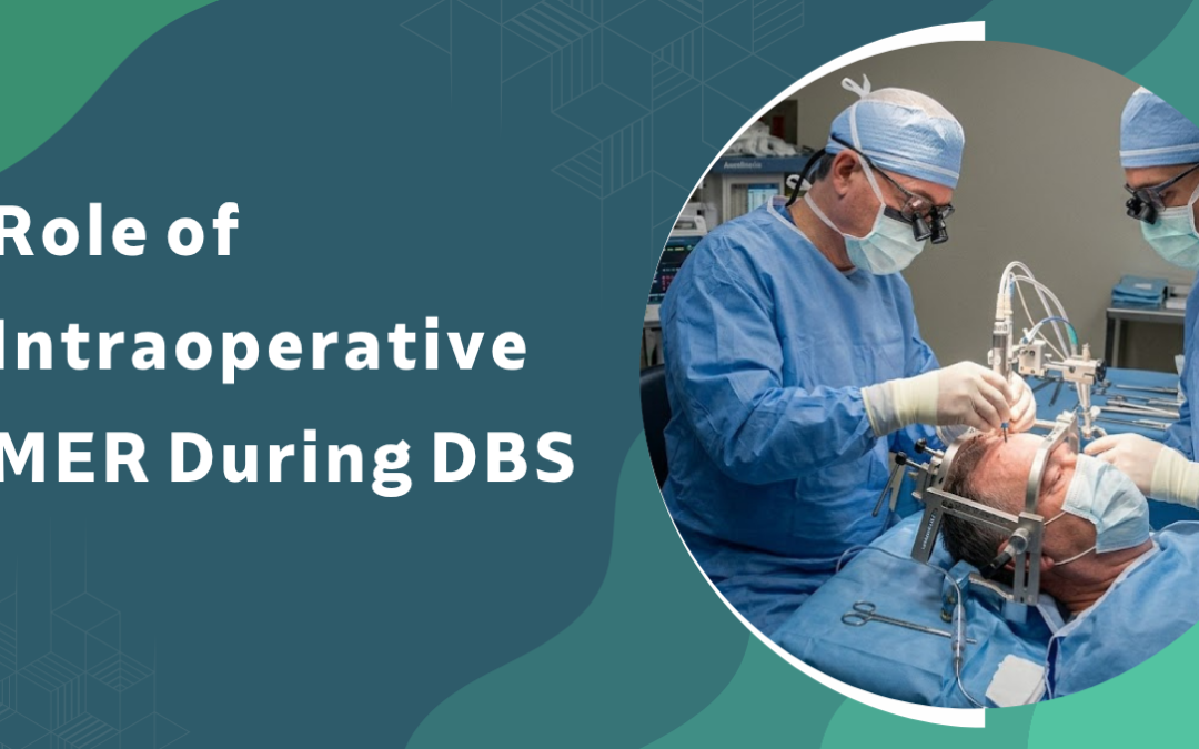
Intraoperative microelectrode recording, or MER, acts as a real-time functional mapping tool during deep brain stimulation surgery that confirms electrode placement within the precise target neurons controlling movement, refines lead position based on live single-unit firing patterns, and corrects for the imaging inaccuracies that frame-based or MRI coordinates alone cannot resolve. It records neuronal activity in sub-millimetre steps along the planned trajectory, distinguishes the motor territory of the subthalamic nucleus from neighbouring structures, and guides the surgeon toward the trajectory most likely to deliver durable symptom control. Imaging plans the route. MER verifies the destination.
According to Dr. Gurneet Singh Sawhney, widely regarded as the best neurosurgeon in Mumbai, Image-based coordinates get the electrode close to the target. MER tells us when we’re inside it, and that’s a different question entirely.
Worried the electrode might land just outside the therapeutic sweet spot?
How Does MER Work Inside the Operating Room?
MER captures neuronal signatures along the surgical track and helps the team interpret them while the electrode is still moving. The patient is usually awake so clinical responses can be tested alongside the recordings. Each pass produces firing patterns that correspond to specific brain regions.
- Signal capture: A high-impedance microelectrode advances in sub-millimetre steps, pausing to record spontaneous neuronal activity at every depth.
- Pattern reading: Different nuclei produce characteristic firing rates and burst patterns, and the team uses these to identify entry into and exit from the target.
- Beta activity: Background activity in the low beta range (around 13 to 20 Hz) maps strongly to the dorsolateral motor STN, which is the region most responsive to stimulation.
- Trajectory choice: When multiple parallel tracks are recorded simultaneously in a Ben’s gun arrangement, the trajectory with the longest and clearest target signal is usually selected for the permanent lead.
The recording itself takes minutes per side. The interpretation is what carries the weight of the decision. For a closer look at the procedure context, see DBS surgery in Mumbai.
Why Does MER Matter for Surgical Outcomes?
MER feeds two decisions at once: whether the electrode is in the right structure, and whether the planned trajectory should be adjusted before the permanent lead goes in. Both decisions shape long-term symptom control.
- Repositioning: Published series report that MER and intraoperative test stimulation lead to a change from the imaging-based plan in a meaningful share of trajectories, especially in earlier years of a programme.
- Sweet spot: A longer recorded target signal generally correlates with the active contact sitting inside the motor territory of the nucleus, which links to better motor improvement at one year.
- Side-effect avoidance: Test stimulation paired with MER lets the team detect capsular or oculomotor side effects before committing to the final lead location, reducing the chance of speech or visual issues later. For programming-stage adjustments, neurostimulator management becomes far cleaner when the lead is well placed.
- Confidence in asleep cases: Newer directional lead technology has narrowed the gap, but in atypical anatomy or revision surgery, MER still resolves uncertainty that imaging alone can’t.
The benefit isn’t uniform across every patient. It’s largest where the anatomy is borderline or the imaging target is ambiguous.
Comparison: Awake DBS With MER vs Asleep DBS Without MER
|
Factor |
Awake DBS with MER |
Asleep DBS without MER |
|
Target verification |
Electrophysiological + clinical |
Imaging only |
|
Patient cooperation |
Required |
Not required |
|
Operative time |
Longer |
Shorter |
|
Useful in atypical anatomy |
Strongly |
Limited |
Why Choose Dr. Gurneet Singh Sawhney?
Dr. Gurneet Singh Sawhney is a senior consultant neurosurgeon with fellowship training in functional neurosurgery and over a decade of experience in movement disorder surgery, including subthalamic and pallidal DBS for Parkinson’s disease, dystonia, and tremor.
Patients are evaluated with a structured pre-operative protocol, awake intraoperative testing where appropriate, and long-term programming follow-up. Lead position is verified, not assumed.
FAQ's
Is MER required for every DBS surgery?
No. It’s standard for awake STN-DBS but optional in many asleep, image-guided protocols.
Does MER make the surgery longer?
Yes, typically by one to two hours per side because of step-wise recording and testing.
Is MER painful for the patient?
The brain itself has no pain receptors, so recording is not painful, only the scalp incision is anaesthetised.
Can MER find the wrong target?
It can mislead in rare cases, which is why clinical test stimulation is always used alongside it.