

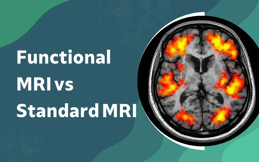
Standard MRI and fMRI are complementary, not competing. MRI shows the tumour’s size, location, and anatomy, while fMRI maps critical functions such as movement, speech, and memory. For tumours near eloquent brain areas, both are essential for safe surgical planning.
According to Dr. Gurneet Singh Sawhney, a leading neurosurgeon in mumbai, surgeons who skip fMRI before operating near eloquent cortex are making the planning decision on anatomy alone. That is not enough. The functional map changes where the margins are drawn.
Brain tumour near a motor or language area with surgery being discussed?
What Does Each Type of MRI Show?
Same machine. Completely different outputs. One is anatomy. The other is activity.
Standard MRI: tumour size, location, signal, relationship to surrounding structures. T1, T2, FLAIR, DWI, contrast. Each sequence adds a different layer of anatomical detail. Always first.
Functional MRI: BOLD signal changes while the patient performs tasks inside the scanner. Hand movement. Naming objects. Reading. The active regions light up and get mapped against the anatomy.
What fMRI cannot do: no tumour detection, no haematoma, no pathology at all. Without the structural MRI underneath it, the functional map floats without context.
Task dependency: patient must be awake, cooperative and able to perform tasks reliably inside the scanner. Acutely unwell patients, confused patients, sedated patients none of them can complete the protocol.
Standard MRI leads every brain tumour workup. Brain tumor surgery planning adds fMRI specifically when functional margins need to be defined alongside anatomical ones.
When Is Functional MRI Specifically Needed?
Not routine. Not for every brain tumour. Ordered when the tumour’s location raises a specific question that structural imaging cannot answer.
Tumour near motor cortex: fMRI identifies hand, face and leg motor representations precisely, so the surgical corridor can be planned around them rather than through them.
Tumour near language areas: left hemisphere lesions near Broca’s or Wernicke’s area need language mapping before surgery. That mapping guides the aphasia risk conversation and the awake craniotomy decision.
Epilepsy surgery planning: fMRI lateralises language and maps memory-critical regions before temporal lobe resection. Complements the EEG and neuropsychological workup.
Intraoperative integration: fMRI data loads directly into the neuronavigation system. The functional overlay runs in real time during surgery alongside the anatomical image.
A pre-operative planning tool. Not a diagnostic one. This guide on recovery after brain tumor surgery covers what post-operative function looks like when functional mapping has shaped what the surgeon removed and what was left alone.
Why Choose Dr. Gurneet Singh Sawhney?
Dr. Gurneet Singh Sawhney trained in Japan. Over 18 years incorporating functional MRI into pre-operative planning for brain tumour and epilepsy surgery. Awake craniotomy for eloquent cortex tumours is part of that practice.
Most patients referred for brain surgery near a motor or language area have never heard of fMRI. They arrive with a structural scan and a diagnosis. The functional map is what comes next — and getting it interpreted by the surgeon who will use it in theatre is what makes it clinically useful rather than just another report.
Frequently Asked Questions
Is functional MRI the same as a standard MRI?
No, standard MRI shows anatomy; functional MRI maps brain activity during specific tasks.
When is functional MRI used before brain surgery?
When a tumour is near motor or language areas and safe resection margins need defining.
Can functional MRI replace awake craniotomy?
No, fMRI informs planning but intraoperative cortical mapping during awake craniotomy remains the gold standard.
Does everyone with a brain tumour need a functional MRI?
No, only when the tumour is near eloquent cortex and surgical resection is being planned.
Disclaimer: The information shared in this content is for educational purposes only and not for promotional use.