
A brain aneurysm is a focal bulge in the wall of a cerebral artery from structural weakness, most commonly at arterial bifurcations in the circle of Willis. Rupture causes subarachnoid haemorrhage with 30-day mortality exceeding 40 percent. Recognising warning signs before that happens is what separates planned intervention from emergency management.
According to Dr. Gurneet Singh Sawhney, Neurosurgeon in Mumbai, the thunderclap headache patients describe as the worst headache of their life is a ruptured aneurysm until proven otherwise it requires emergency CT and neurosurgical assessment the same day, not the next morning.
What Is a Brain Aneurysm and How Does It Develop?
Brain aneurysms form at points of arterial wall weakness where haemodynamic stress concentrates. Most patients have no idea the lesion exists until it bleeds or gets found on imaging done for something else entirely.
- Saccular aneurysms: The most common type, arising as rounded outpouchings at arterial bifurcations in the circle of Willis. They account for over 90 percent of all intracranial aneurysms and carry the highest rupture risk of all aneurysm types.
- Fusiform aneurysms: Caused by diffuse arterial wall weakening producing circumferential dilation rather than a discrete sac. These less commonly rupture but can produce symptoms through mass effect on adjacent cranial nerves before any bleed occurs.
- Risk factors: Hypertension, smoking, family history of intracranial aneurysm, polycystic kidney disease, and connective tissue disorders are established risk factors. Aneurysm size above seven millimetres and posterior circulation location carry significantly higher rupture risk than small anterior circulation lesions.
- Incidental detection: Most unruptured aneurysms are found on MRI or CT angiography done for unrelated reasons. But incidental detection does not mean the lesion is safe it means the rupture has not happened yet, and that is precisely the window for planned treatment before a catastrophe occurs.
Brain aneurysm diagnosis requires CT angiography or MR angiography for initial characterisation and catheter cerebral angiography for definitive treatment planning.
Explore functional neurosurgery in Mumbai for brain aneurysm evaluation and treatment planning at Fortis Hospital Mulund West.
What Are the Warning Signs Before a Brain Aneurysm Ruptures?
Warning signs appear in a proportion of patients days to weeks before a major bleed. Acting on them immediately is the window that prevents the catastrophic haemorrhage that follows. Missing them or dismissing them closes that window permanently.
- Sentinel headache: A sudden severe headache distinctly different from anything the patient has experienced before, occurring days to weeks before major rupture, is a sentinel bleed from a small aneurysmal leak. Because it resolves spontaneously, most patients dismiss it. That dismissal costs them the only opportunity for intervention before a catastrophic rupture occurs.
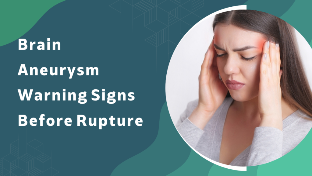
- Thunderclap headache: Instantaneous onset severe headache reaching maximum intensity within seconds the worst headache of the patient’s life is subarachnoid haemorrhage until proven otherwise. CT brain within six hours has sensitivity above 98 percent. It is mandatory the same day. Not tomorrow.
- Third nerve palsy: Sudden onset ptosis, dilated unreactive pupil, and ophthalmoplegia indicates posterior communicating artery aneurysm compressing the third cranial nerve. This is a neurosurgical emergency. Same-day CT angiography and urgent intervention before rupture not observation and review in a week.
- Visual disturbance and focal symptoms: Diplopia, transient visual loss, and focal neurological symptoms in a patient with a known or suspected aneurysm indicate enlargement or sentinel leak. Immediate imaging and neurosurgical review is required without delay because the alternative is waiting for the rupture to make the decision for everyone.
Families who have read about whether neurological problems can exist with normal scans understand why a normal CT does not exclude subarachnoid haemorrhage and lumbar puncture is required when clinical suspicion remains after negative imaging.
Why Choose Dr.Gurneet Singh Sawhney?
Dr. Gurneet Singh Sawhney completed dedicated fellowships in functional neurosurgery under Prof. Taira at Tokyo Women’s Medical University and epilepsy surgery under Prof. Sugano at Juntendo University, both high-volume academic centres where cerebrovascular neurosurgical cases including aneurysm clipping and endovascular management formed a structured part of the caseload. At Fortis Hospital Mulund West, brain aneurysm cases receive CT angiography, catheter angiography where indicated, and direct treatment planning before any management decision is finalised.
Patients with a diagnosed brain aneurysm receive a direct assessment covering size, location, rupture risk, treatment options, and what realistic outcomes look like for that specific aneurysm. Not population averages. A recommendation based on objective imaging and clinical findings for that individual case alone.
FAQ's
What is the most important warning sign of brain aneurysm rupture?
A thunderclap headache of instantaneous onset reaching maximum intensity within seconds requires same-day emergency CT brain assessment without exception.
What is a sentinel headache and why does it matter?
A sentinel headache is a sudden severe headache from a small aneurysmal leak days to weeks before major rupture, frequently dismissed because it resolves spontaneously.
What causes a brain aneurysm to form?
Hypertension, smoking, family history, polycystic kidney disease, and connective tissue disorders are established risk factors for intracranial aneurysm formation and growth.
Is an unruptured brain aneurysm always treated with surgery?
Treatment decisions depend on size, location, patient age, and rupture risk estimate not all unruptured aneurysms require immediate intervention.
References
- National Institute of Neurological Disorders and Stroke. Neurological Diagnostic Tests and Procedures. NINDS, NIH.
2. Vlak MH, et al. Prevalence of Unruptured Intracranial Aneurysms. PubMed Central, NCBI.