
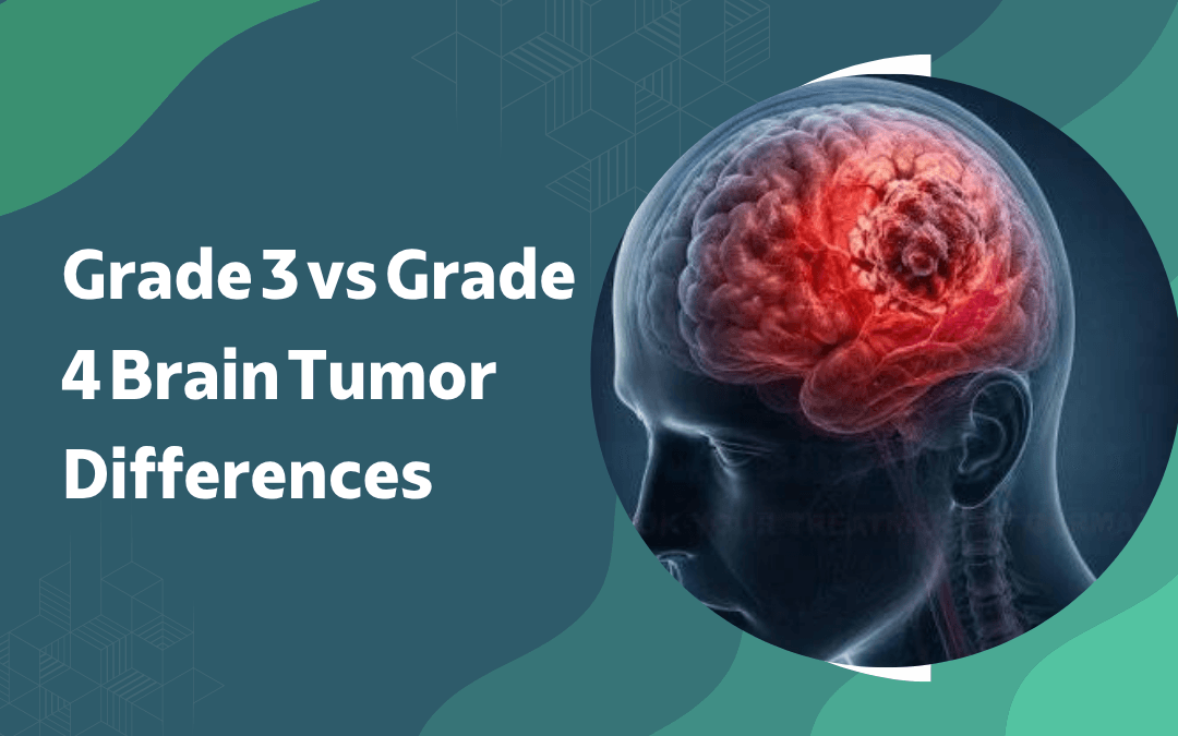
Both grade 3 and grade 4 brain tumors are classified as high-grade malignancies under the WHO system, meaning both grow rapidly, infiltrate surrounding tissue, and require aggressive multimodal treatment but they aren’t the same disease. Grade 3 tumors, such as anaplastic astrocytoma, show active cell division and early vascular changes without the necrotic core that defines grade 4. Grade 4 glioblastoma adds microvascular proliferation and central necrosis to that picture, producing a more aggressive growth pattern and a shorter median survival. The distinction shapes every treatment decision from surgical planning through adjuvant therapy.
According to Dr. Gurneet Singh Sawhney, best neurosurgeon in Mumbai, “Grade 3 and grade 4 aren’t just numbers on a pathology report they determine how urgently surgery happens, how radiation is dosed, and what chemotherapy the patient qualifies for.”
Diagnosed with a high-grade glioma and uncertain about the next steps?
How Do Grade 3 and Grade 4 Tumors Differ in Biology and Diagnosis?
The pathological gap between grade 3 and grade 4 is specific and determines treatment eligibility, not just prognosis.
Cell behaviour: Grade 3 tumors show nuclear atypia and active mitosis — cells dividing faster than normal tissue, while grade 4 adds pseudopalisading necrosis, where tumor cells arrange around dead tissue, a pattern specific to glioblastoma and absent in grade 3.
Molecular markers: IDH mutation status and MGMT promoter methylation now define grade as much as histology does, with IDH-mutant grade 3 tumors carrying better outcomes than IDH-wildtype grade 4 tumors, even when the imaging looks similar.
Imaging features: Grade 4 glioblastoma typically shows a ring-enhancing lesion with central necrosis on contrast MRI, a pattern grade 3 tumors don’t produce, though imaging alone can’t confirm grade, and tissue biopsy remains the diagnostic standard.
Onset speed: Grade 3 tumors can evolve from lower-grade precursors over months to years, whereas grade 4 glioblastoma frequently presents de novo with rapid symptom onset over weeks, often leaving little time between first symptom and diagnosis.
Tissue diagnosis with full molecular profiling is the only way to accurately separate grade 3 from grade 4, and treatment planning shouldn’t begin without it. Surgical resection in both cases follows the same foundational principles used across brain surgery for infiltrative malignancies.
How Does Treatment Differ Between Grade 3 and Grade 4 Tumors?
Both grades require surgery, radiation, and chemotherapy but dosing, sequencing, and eligibility criteria diverge significantly.
Surgical goal: Maximum safe resection is the target in both grades, but grade 4 glioblastoma demands faster surgical turnaround given its growth rate, and intraoperative fluorescence-guided resection with 5-ALA is more commonly indicated to push resection extent in GBM than in grade 3 tumors.
Radiation dosing: Grade 4 glioblastoma is treated with 60 Gy over 30 fractions concurrently with temozolomide as standard of care, while grade 3 anaplastic gliomas typically receive 59.4 Gy over a longer fractionation schedule with chemotherapy sequencing determined by IDH and 1p/19q status.
Chemotherapy eligibility: MGMT promoter methylation in grade 4 predicts response to temozolomide and directly influences adjuvant treatment selection a molecular variable that functional neurosurgery planning teams factor in when mapping eloquent cortex before resection, since preserving function extends the window for adjuvant therapy to work.
Prognosis gap: Median survival for IDH-mutant grade 3 anaplastic astrocytoma runs several years with optimal treatment, while grade 4 glioblastoma carries a median survival of 14 to 18 months on standard therapy a gap wide enough to make accurate grading one of the most consequential steps in the diagnostic workup.
Recurrence is expected in both grades and surveillance imaging every two to three months post-treatment is standard practice regardless of initial response. For context on what separates high-grade from lower-grade malignancies at the broad diagnostic level, see benign vs malignant brain tumors.
Why Choose Dr. Gurneet Singh Sawhney?
Dr. Gurneet Singh Sawhney holds MBBS, MS (General Surgery), and MCh (Neurosurgery) — ranking first in the MCh university examination with fellowship training in Functional Neurosurgery and Epilepsy Surgery from Japan, specialised expertise in Neuro-oncology and Neuroendoscopy, and over 18 years of overall surgical experience managing high-grade glioma cases across all stages.
His approach to grade 3 and grade 4 tumors integrates intraoperative mapping, fluorescence-guided resection where indicated, and molecular profiling before adjuvant treatment is planned because surgical outcome and treatment eligibility are decided together, not in sequence.
Frequently Asked Questions
Can a grade 3 tumor progress to grade 4?
Yes, grade 3 gliomas can transform to glioblastoma over time without treatment.
Is grade 3 brain tumor always fatal?
Not always do IDH-mutant grade 3 tumors carry significantly better long-term outcomes than grade 4.
Does grade determine which chemotherapy a patient receives?
Yes, molecular grade and MGMT status directly determine chemotherapy eligibility and sequencing.
Can imaging alone confirm a brain tumor's grade?
No, tissue biopsy with molecular profiling is required to confirm grade accurately.
Disclaimer: The information shared in this content is for educational purposes only and not for promotional use.