
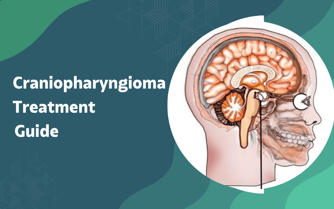
Craniopharyngioma is a benign, slow-growing tumor that develops near the pituitary gland and hypothalamus, arising from remnants of embryonic tissue left over from fetal development. It doesn’t invade surrounding brain tissue the way malignant tumors do, but its location directly alongside structures that regulate hormones, vision, and fluid balance makes it clinically complex. Children between five and fourteen and adults in their fifties and sixties are most commonly affected. Benign doesn’t mean harmless here. The tumor’s proximity to critical anatomy is what drives the difficulty.
According to Dr. Gurneet Singh Sawhney, best neurosurgeon in Mumbai, “A craniopharyngioma’s grade on imaging tells you almost nothing what determines the outcome is how much of the hypothalamus and optic structures the tumor has involved before surgery begins.”
Child showing unexplained vision loss, growth delay, or persistent headaches?
What Symptoms Does a Craniopharyngioma Typically Cause?
Symptom onset is gradual, often spanning months before a clinical picture becomes clear enough to prompt imaging.
Vision loss: The tumor compresses the optic chiasm from below, producing bitemporal hemianopia loss of the outer visual fields in both eyes which patients and families frequently attribute to a refractive problem before a neurological cause is considered.
Hormonal disruption: Pressure on the pituitary and hypothalamus disrupts growth hormone, thyroid, and antidiuretic hormone production, causing short stature in children, delayed puberty, diabetes insipidus, and weight gain that doesn’t respond to dietary changes.
Raised pressure: As the tumor enlarges it blocks cerebrospinal fluid drainage, producing classic raised intracranial pressure symptoms morning headache, nausea, and vomiting that worsen progressively rather than fluctuating the way tension headaches do.
Cognitive changes: Hypothalamic involvement can impair memory, attention, and emotional regulation, particularly in children, sometimes presenting as academic decline or behavioural problems before any symptoms surface.
Symptoms don’t always arrive together and the full picture can take a year or more to develop, which is why craniopharyngioma is frequently misdiagnosed on first presentation. Surgical access to this region follows the same planning frameworks used in brain surgery for deep midline lesions.
How Is a Craniopharyngioma Surgically and Medically Treated?
Treatment strategy is determined by tumor size, degree of hypothalamic involvement, and the patient’s age; there’s no single standard approach.
Endoscopic removal: For tumors with primarily sellar and suprasellar extension, the endoscopic endonasal transsphenoidal route allows access through the nasal passage without a skull opening, reducing operative trauma and recovery time significantly in appropriately selected cases.
Craniotomy: Tumors extending laterally or involving the third ventricle require an open craniotomy, with the surgical corridor chosen based on which approach preserves the most hypothalamic tissue functional neurosurgery principles directly inform how the surgical team maps and protects adjacent structures throughout.
Radiation: When complete removal would damage the hypothalamus too severely, planned subtotal resection followed by stereotactic radiosurgery or fractionated radiotherapy controls residual tumor while limiting injury to surrounding tissue a strategy used increasingly in children to protect developing endocrine function.
Hormone replacement: Surgery almost always disrupts pituitary function to some degree, making lifelong hormone replacement therapy covering thyroid, adrenal, and growth hormone axes a standard part of post-operative management rather than an exceptional complication.
Recurrence remains a genuine risk even after complete resection, and long-term follow-up imaging every one to two years is standard practice regardless of the surgical approach taken. For broader context on brain tumor warning signs that precede diagnosis, see brain tumor warning signs.
Why Choose Dr. Gurneet Singh Sawhney?
Dr. Gurneet Singh Sawhney holds MBBS, MS (General Surgery), and MCh (Neurosurgery) qualifications ranking first in the MCh university examination with fellowship training in Functional Neurosurgery and Epilepsy Surgery from Japan, specialised expertise in Neuroendoscopy, and over 18 years of overall surgical experience managing complex brain and skull-base cases.
His surgical approach to deep midline tumors prioritises hypothalamic preservation and functional outcome over maximal resection, with post-operative hormone management coordinated as part of the treatment plan from the outset not as an afterthought.
Frequently Asked Questions
Is a craniopharyngioma cancerous?
No, it’s classified as benign but causes serious harm through compression of nearby structures.
Can craniopharyngioma recur after complete removal?
Yes, recurrence is possible even after gross total resection, requiring long-term imaging follow-up.
Does surgery always affect pituitary function?
In most cases yes, hormone replacement therapy is required post-operatively.
Is craniopharyngioma more common in children or adults?
It occurs in two age peaks children aged five to fourteen and adults in their fifties.
Disclaimer: The information shared in this content is for educational purposes only and not for promotional use.