
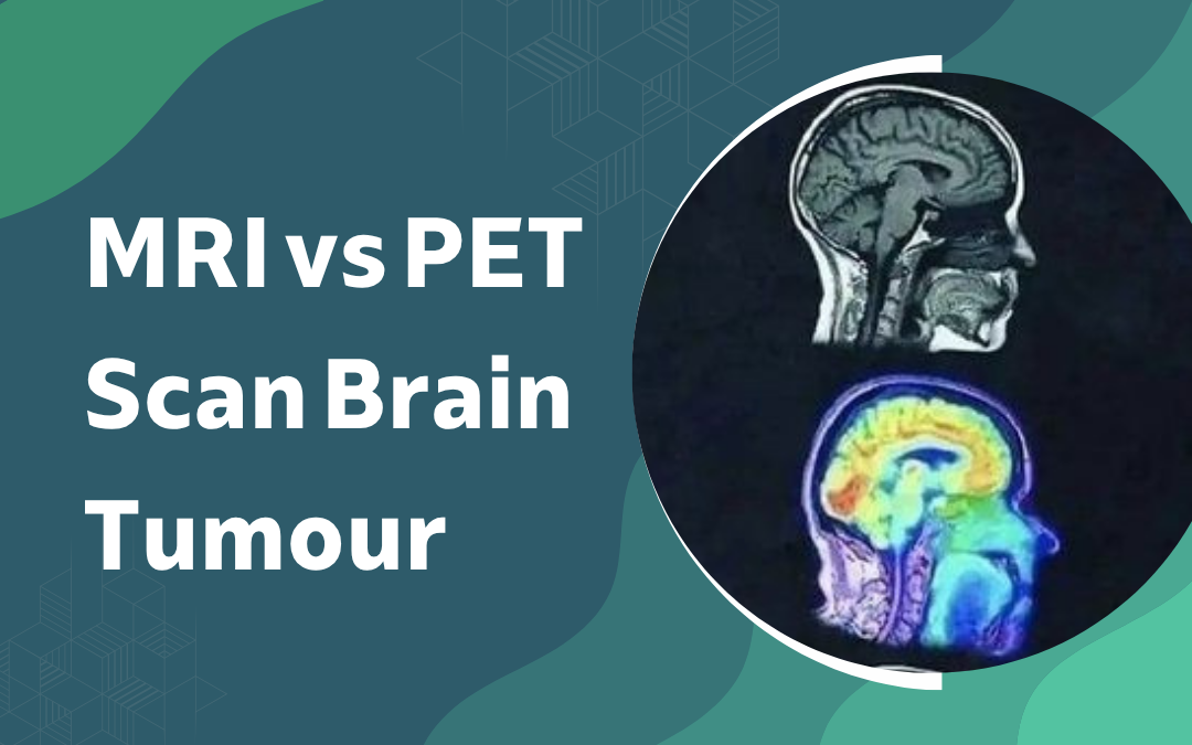
Neither is universally better. MRI is the first investigation and the one that drives surgical planning. PET follows when the question shifts from where the tumour is to how metabolically active it is, or whether a change on post-treatment imaging represents recurrence or treatment effect. For most patients the two are not competing options. They work in sequence.
According to Dr. Gurneet Singh Sawhney, a leading neurosurgeon in Mumbai, the question is not which scan is better, it is what question needs answering. MRI tells you where the tumour is and what it looks like. PET tells you how metabolically active it is. For planning surgery or deciding whether a change on MRI is recurrence or radiation damage, both pieces of information are often needed.
Brain tumour diagnosis in progress and unsure what the imaging findings mean for treatment?
Why Is MRI Always the First Investigation?
MRI is the cornerstone of every brain tumour workup from the start. Anatomy, structure, surgical planning none of that comes from PET.
Structure: MRI with gadolinium contrast maps tumour size, location and relationship to eloquent cortex, the roadmap the surgeon works from before and during the operation
Staging: FLAIR, DWI and perfusion sequences reveal infiltration extent and restricted diffusion that contrast enhancement alone misses, directly informing grading and margin decisions
Surgery: intraoperative MRI and functional mapping run entirely on MRI data, because the operating theatre needs precise anatomy, not metabolic signal
Surveillance: post-treatment follow-up is MRI by default, monitoring for recurrence, tracking response and identifying complications like radiation necrosis before they become clinical problems
For diagnosis, characterisation and surgical planning, MRI is the irreplaceable standard. That is where brain tumor surgery begins and where it stays.
When Does a PET Scan Change the Clinical Picture?
PET answers the questions MRI raises but cannot resolve. Most of that work happens after treatment, when the imaging picture becomes harder to interpret.
Recurrence vs radiation necrosis: a new enhancing lesion on post-treatment MRI could be either, and PET measures metabolic activity at the site to differentiate them, which directly changes the clinical decision
Grading: amino acid tracers concentrate in high-grade tumour, and PET can direct biopsy to the most metabolically active region rather than leaving grade to sampling chance
Extent: infiltrating tumours often spread beyond their MRI-visible margins, and PET maps this metabolic footprint, relevant for radiotherapy planning and understanding true disease burden
Treatment response: chemoradiation response can appear metabolically on PET before any anatomical change is visible on MRI, allowing earlier and more informed decision-making
When the clinical question outgrows what anatomy alone can answer, PET fills the gap. This guide on benign vs malignant brain tumours covers how imaging findings feed into the malignancy assessment.
Why Choose Dr. Gurneet Singh Sawhney?
Dr. Gurneet Singh Sawhney trained in Japan, has spent over 18 years operating on brain tumours and reads both MRI and PET in the context of planning and post-treatment decisions. The imaging doesn’t sit in a radiology report. It drives the surgical plan.
Reading brain tumour imaging in the context of a surgical plan is a different skill from reading it in isolation. Dr. Sawhney’s practice integrates both MRI and PET findings into pre-operative planning and post-treatment decision-making, so the imaging review at the first consultation produces a clinical plan, not just a report summary.
Frequently Asked Questions
Is MRI or PET scan done first for a suspected brain tumour?
MRI with contrast is always the first investigation for a suspected brain tumour.
Can a PET scan detect brain tumours better than MRI?
No, PET adds metabolic information but MRI remains superior for anatomical detail and diagnosis.
When is a PET scan used instead of MRI for brain tumours?
PET is not used instead of MRI; it is added when metabolic information is needed.
Can a PET scan distinguish tumour recurrence from radiation damage?
Yes, PET measures metabolic activity and can reliably differentiate recurrence from radiation necrosis.
Disclaimer: The information shared in this content is for educational purposes only and not for promotional use.