
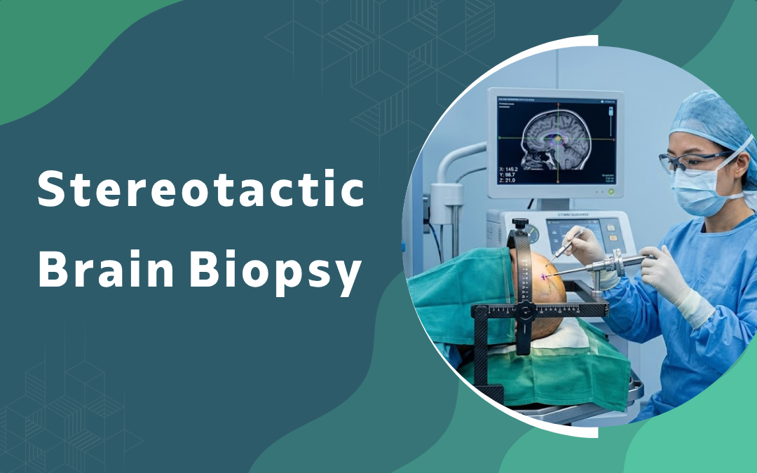
A stereotactic brain biopsy is a minimally invasive neurosurgical procedure that uses 3D imaging from CT or MRI scans and computer-guided navigation to pass a thin needle through a small skull opening and sample tissue from deep-seated or hard-to-reach brain lesions. It’s primarily required for diagnosing tumors, infections like abscesses, inflammatory conditions, and certain neurodegenerative diseases when non-invasive imaging stays inconclusive.
According to Dr. Gurneet Singh Sawhney, Neurosurgeon in Bangalore, When the MRI shows something off but won’t tell us what, that’s when a biopsy stops being optional. We need cells, not pictures.
When does a doctor recommend stereotactic brain biopsy?
The decision rests on two clinical factors, the exact location of the lesion and the diagnostic gaps that imaging alone cannot address.
- Deep lesions: Some tumors hide in places you really don’t want to open up, the brainstem, thalamus, deep down in the basal ganglia, and a needle through a tiny hole in the skull is just smarter than going in big.
- MRI confusion: A scan can show you a mass and still leave the room split, is it a tumor, is it an abscess, is it something autoimmune, and you can’t keep guessing forever, the tissue has to settle it.
- Multiple spots: When small lesions are scattered all over both sides, you don’t biopsy each one, you pick the one that’s easiest to reach safely and trust pathology to do the rest.
- Suspected lymphoma: Brain lymphoma is funny that way, you don’t cut it out, you treat it with chemo and radiation, so confirming it on biopsy first is non-negotiable, surgery would actually be the wrong move.
If your scan came back with something nobody could fully explain, getting a brain tumour evaluation is honestly the cleanest next step.
How is the procedure actually done?
Stereotactic Brain Biopsy Procedure
| Step | Stage | Details |
|---|---|---|
| Step 1 | Frame Setup | Stereotactic frame fixed plus MRI mapping |
| Step 2 | Burr Hole | Small skull opening less than one centimetre |
| Step 3 | Sample Retrieval | Two or three tiny samples plus frozen section |
| Step 4 | Recovery | One night stay then home next morning |
Why Choose Dr.Gurneet Singh Sawhney?
Dr. Gurneet Singh Sawhney is a senior neurosurgeon with 15+ years of experience in complex brain and spine cases, including frame-based and frameless stereotactic procedures across teaching and corporate hospital settings.
What patients keep saying is that the conversation before the biopsy was the part that helped most. They knew exactly where the needle was going, what the risk numbers actually meant, and what the report would decide on the other side. No surprises in the OR.
FAQ's
What is the success rate of stereotactic brain biopsy?
Diagnostic accuracy sits around 95% with experienced surgeons and modern navigation systems.
Is stereotactic brain biopsy painful?
The procedure runs under local anesthesia with sedation, so most people feel only mild pressure.
How long does the biopsy take?
The actual biopsy runs 30 to 60 minutes, full setup with imaging adds another hour.
When will I get the biopsy results?
Preliminary results come within 24 hours, final pathology with typing takes 5 to 7 days.
References
- Stereotactic Biopsy of Brain Lesions — PubMed, NIH
- Brain Tumor Diagnosis Guidelines — National Cancer Institute