
Hydrocephalus is abnormal accumulation of cerebrospinal fluid within the ventricular system, causing raised intracranial pressure. In children it results from obstruction to CSF flow, impaired absorption, or rarely excess production. Surgical intervention is required in the majority of cases the developing brain tolerates raised intracranial pressure poorly and neurological damage from untreated hydrocephalus is cumulative and frequently irreversible.
According to Dr. Gurneet Singh Sawhney, Neurosurgeon in Mumbai, hydrocephalus in children is a neurosurgical condition the question is not whether to treat but which procedure is appropriate for that specific child’s anatomy, aetiology, and age at presentation.
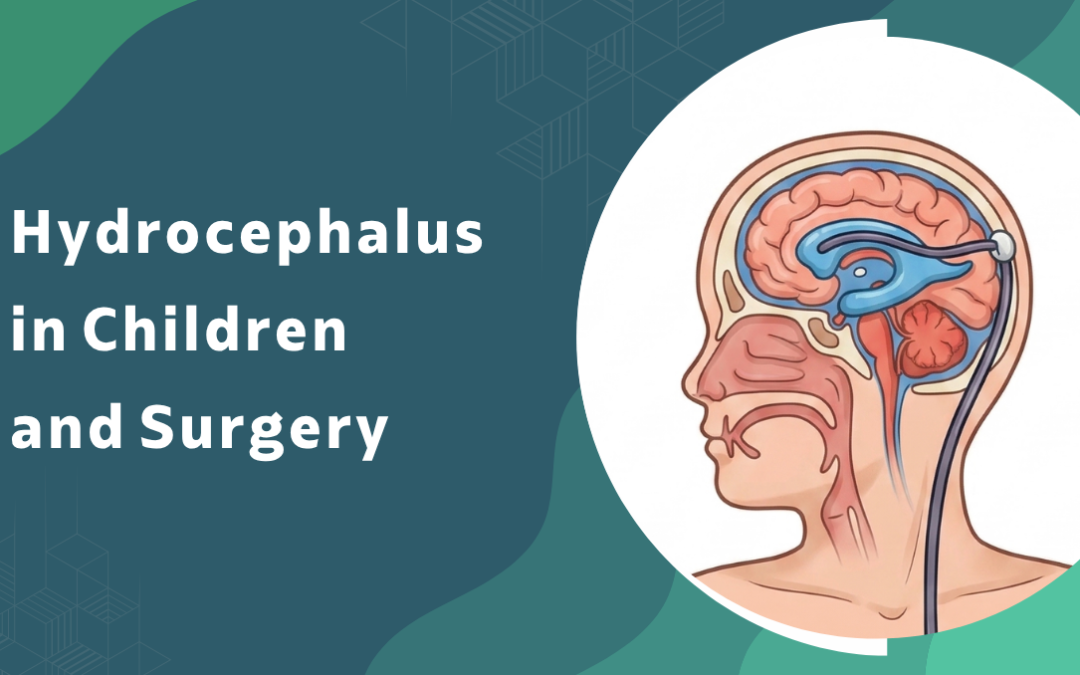
What Is Hydrocephalus and How Does It Present in Children?
Presentation differs significantly by age at onset and rate of CSF accumulation. Missing the signs early costs neurological function that timely surgical decompression would have preserved.
- Presentation in infants: Rapidly enlarging head circumference crossing centile lines, bulging anterior fontanelle, sunset sign of the eyes, irritability, and poor feeding are the cardinal features. Because the unfused skull accommodates expanding ventricles initially without immediate pressure signs, regular head circumference monitoring is what catches this before the window closes.
- Presentation in older children: Headache on waking, vomiting, blurred vision from papilloedema, deteriorating school performance, and gait disturbance dominate in children with fused skull sutures. And progressive visual loss from chronic papilloedema is irreversible which makes delayed diagnosis in this age group particularly consequential.
- Causes in paediatric cases: Congenital aqueduct stenosis, post-haemorrhagic hydrocephalus in premature infants, posterior fossa tumours, spina bifida-associated Chiari malformation, and post-infectious hydrocephalus from neonatal meningitis account for the majority of cases. Because aetiology determines both surgical approach and long-term prognosis, identifying the cause is not separate from planning treatment.
- Investigation before surgery: MRI brain and spine defines ventricular anatomy, identifies the obstruction site, and determines whether hydrocephalus is communicating or non-communicating. Because this distinction directly determines whether endoscopic third ventriculostomy or VP shunting is appropriate, imaging quality and sequence selection matter before any surgical planning begins.
Paediatric hydrocephalus requires neurosurgical evaluation at diagnosis. Monitoring without treatment in a symptomatic child with documented ventricular enlargement is not appropriate management.
Explore functional neurosurgery in Mumbai and paediatric neurosurgical evaluation at Fortis Hospital Mulund West.
When Does Hydrocephalus in Children Require Surgery?
The majority of symptomatic paediatric hydrocephalus requires surgical treatment. Procedure selection depends on obstruction site, patient age, and ventricular anatomy on imaging.
- Symptomatic raised intracranial pressure: Documented ventricular enlargement with clinical features of raised pressure is a surgical indication without exception. Waiting for further neurological deterioration in a child with confirmed hydrocephalus and symptoms is not a defensible clinical position.
- Endoscopic third ventriculostomy: ETV creates a bypass through the floor of the third ventricle, allowing CSF to drain into the basal cisterns and avoiding a shunt entirely. Because it carries no implant-related complications, ETV is the preferred procedure for non-communicating obstructive hydrocephalus in children over six months with appropriate anatomy confirmed on MRI.
- Ventriculoperitoneal shunt: VP shunting is indicated when ETV is not feasible due to communicating hydrocephalus, failed ETV, or unfavourable ventricular anatomy. A programmable valve allows non-invasive pressure adjustment post-implantation particularly important in young children where optimal settings change with growth and posture over time.
- Post-haemorrhagic and post-infectious hydrocephalus: Premature infants and children with post-meningitic hydrocephalus frequently require temporary CSF diversion through a ventricular reservoir before definitive shunting when CSF protein levels are too high for a permanent shunt to function reliably.
Earlier intervention consistently produces better neurodevelopmental outcomes than delayed treatment. Cortical thinning from prolonged ventricular enlargement does not fully recover after decompression timing matters more than most families are told.
Families who have read about whether neurological problems can exist with normal scans understand why MRI protocol selection is essential in paediatric hydrocephalus planning rather than CT alone.
Why Choose Dr.Gurneet Singh Sawhney?
Dr. Gurneet Singh Sawhney completed dedicated fellowships in functional neurosurgery under Prof. Taira at Tokyo Women’s Medical University and epilepsy surgery under Prof. Sugano at Juntendo University, both high-volume academic centres where paediatric neurosurgical cases including hydrocephalus management formed a structured part of the caseload. At Fortis Hospital Mulund West, paediatric hydrocephalus cases receive MRI evaluation, aetiology assessment, and a direct recommendation covering procedure selection and surgical timing before any decision is finalised.
Families presenting with a child diagnosed with hydrocephalus receive a structured assessment covering cause, ventricular anatomy, ETV candidacy, shunt selection where indicated, and what realistic neurodevelopmental outcomes look like for that specific case. The recommendation is based on objective imaging and clinical findings.
FAQ's
What is hydrocephalus in children and what causes it?
Hydrocephalus is abnormal CSF accumulation in the brain’s ventricular system causing raised intracranial pressure, resulting from obstruction, impaired absorption, or congenital malformation.
When does hydrocephalus in children require surgery?
Symptomatic hydrocephalus with documented ventricular enlargement and raised intracranial pressure features requires surgical treatment without delay in all paediatric cases.
What is endoscopic third ventriculostomy and when is it used?
ETV creates a CSF bypass through the third ventricle floor avoiding a shunt, preferred for non-communicating obstructive hydrocephalus in children over six months with appropriate anatomy.
What is a ventriculoperitoneal shunt and when is it needed?
A VP shunt diverts CSF from the ventricles to the peritoneum and is indicated when ETV is not feasible due to communicating hydrocephalus or unfavourable ventricular anatomy.
References
- National Institute of Neurological Disorders and Stroke. Neurological Diagnostic Tests and Procedures. NINDS, NIH.
2. Kulkarni AV, et al. Endoscopic Third Ventriculostomy versus Shunting in Infants. PubMed Central, NCBI.