

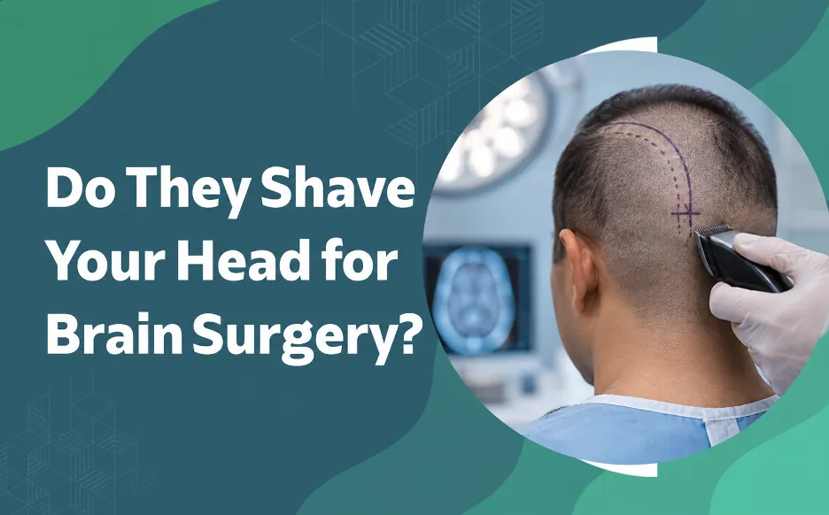
Not necessarily. Whether hair is removed during brain surgery depends on the type of surgery, the incision site, and the technique used. Today, shave-free surgery is increasingly preferred and recommended wherever it can be done safely. Many patients have only a patch or strip of hair clipped, and some have their hair left untouched. Dr. Sawhney has performed brain surgeries without removing hair in suitable cases. When removal is needed, hair is clipped, not razor-shaved, usually in the operating theatre. Regrowth starts within weeks and looks close to normal in about a year. Discuss hair removal with your surgical team before surgery.
“Do they shave your head for brain surgery?” is one of the most common questions patients ask before an operation. It is a sensible one. Hair is closely tied to identity, and the thought of losing it adds to stress at an anxious time. The reality is that practice varies. Modern neurosurgery uses smaller incisions and greater precision, so a complete head shave is no longer necessary, and shave-free surgery is now the approach many surgeons prefer and recommend when the case allows.
According to Dr. Gurneet Singh Sawhney, Neurosurgeon in Mumbai,
“Patients are often relieved to hear that we remove only what the operation needs, and sometimes nothing at all. Shave-free surgery is what we prefer and recommend wherever it is safe to do so. The plan is made before surgery, and we explain it so there are no surprises when you wake up.”
Preparing for Brain Surgery and Have Questions About the Process? Get clear answers before the day. Contact the Clinic to discuss your case.
What to Expect in the First Two Weeks
The period prior to surgery is all about preparation, and hair is one aspect of it. Most of what is done is straightforward and well-planned.
- Pre-operative consultation: Your surgeon discusses the incision point as well as the clipping of hair if needed. It is the right moment to voice worries about your appearance.
- Home hair removal not permitted: Do not shave or cut the hair yourself at home. At-home hair removal tools may result in small cuts which increase the risk of infection.
If you are going to have surgery due to a brain tumor, the planning of your procedure will be carefully done. Find more information on brain tumor surgery and its stages of planning.
Why Is Hair Removal Necessary?
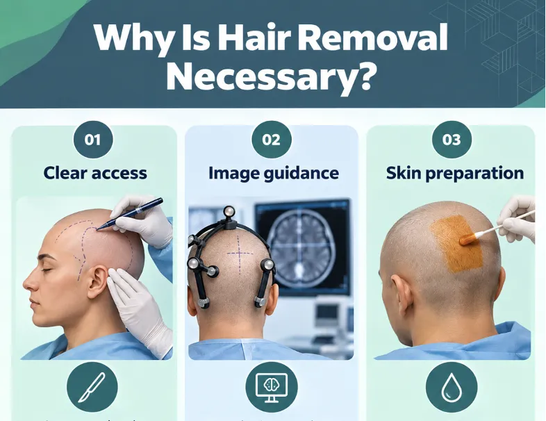
It is not a matter of practice. If your surgeon decides to remove some hair, there are several reasons related to safety and accuracy.
Clear access:
An exposed scalp allows the surgeon to mark and make the incision correctly.
Image guidance:
Many brain procedures require image guidance. Marks and sensors fit better on an exposed scalp.
Skin preparation:
Antiseptic solution gets onto the scalp without hair obstruction.
Shave-Free Brain Surgery: Why It Is Now Preferred and Recommended
Surgery has moved steadily towards leaving hair in place. The World Health Organization’s global guidelines on preventing surgical site infection say hair should either not be removed or, if removal is absolutely necessary, be done only with a clipper, and that shaving is strongly discouraged. The recommendation is set out in the WHO surgical site infection guidance.
A shave-free approach can mean:
- Less visible change after surgery, which many patients find reassuring
- Hair that can help cover the incision once healing allows
Shave-free surgery is not possible in every case. The size and position of the incision, the approach to the brain, and safety all decide whether hair can be left in place. The final decision rests with the operating surgeon.
When Hair Removal is Not Required?
Modern practices of surgeries tend to include fewer hair removals. According to the WHO guidance on prevention of surgical site infection, hair removal should either be avoided or, if removal is absolutely necessary, be done only with a clipper, and that shaving is strongly discouraged. The recommendation is set out in the WHO surgical site infection guidance.
How this applies to brain surgery depends on the approach.
Hair-parting techniques:
In some cases the surgeon parts the hair along the incision line and holds it aside, leaving most of it untouched. Dr. Sawhney has performed brain surgery without shaving the patient’s hair in suitable cases, using this approach.
Small openings:
Procedures through a small burr hole or endoscopic route may need minimal or no clipping.
Awake and functional procedures:
Where the brain’s speech or movement areas are involved, techniques such as awake craniotomy are planned with the patient and team in advance, including how much hair is affected. The same applies to deep brain stimulation for movement disorders.
The final call rests with the operating surgeon, based on safety and what the procedure needs.
Hair Removal by Type of Procedure
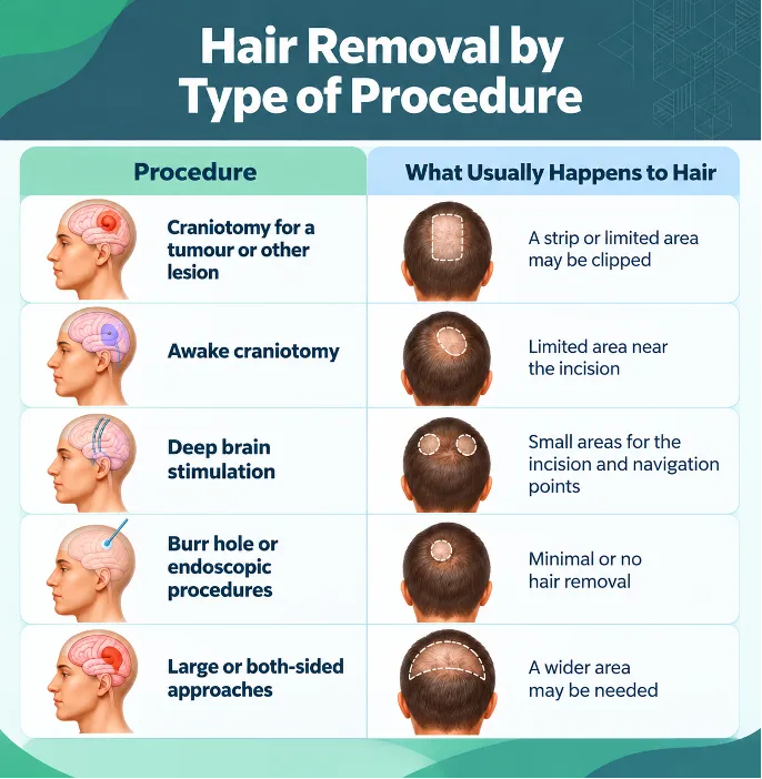
This table is a general guide only. Your surgeon will tell you what applies to your operation.
| Procedure | What Usually Happens to Hair |
| Craniotomy for a tumour or other lesion | A strip or limited area near the incision may be clipped, or hair may be parted in suitable cases |
| Awake craniotomy | Limited area near the incision, planned with the team |
| Deep brain stimulation | Small areas for the incision and frame or navigation points |
| Burr hole or endoscopic procedures | Minimal or no hair removal in many cases |
| Large or both-sided approaches | A wider area may be needed |
Patients considering brain tumor surgery or an awake craniotomy can ask their surgeon how the plan applies to them. For movement disorders, the same discussion applies to deep brain stimulation.
Worried About How You’ll Look After Surgery? Ask About Shave-Free Options. Concerns about hair and scars are valid and worth raising.
How Is Hair Removed Safely?
Hair removal is done in a very careful manner.
- Clipper shaving is done instead of razor shaving: Razor shaving may lead to microscopic skin injuries that increase the risk of infection. That is why clipping is recommended.
- In the operating theatre: Your hair is usually clipped after anesthesia, so you will be unconscious during this process.
- Only hair that needs to be removed: Your surgeon will mark the minimum possible area around your future incision.
- Your beliefs must be respected: Make sure you inform your surgeon or medical staff about your views on hair, if any.
How Long Does Hair Take to Grow Back?
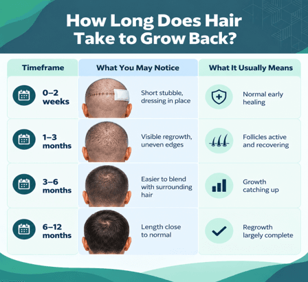
Clipped hair is not lost for good. Follicles remain intact and regrowth follows your usual pace, though age, health, and hair type all play a part. Treat this table as a guide rather than a fixed schedule.
| Timeframe | What You May Notice | What It Usually Means |
| 0–2 weeks | Short stubble, dressing in place | Normal early healing |
| 1–3 months | Visible regrowth, uneven edges | Follicles active and recovering |
| 3–6 months | Easier to blend with surrounding hair | Growth catching up |
| 6–12 months | Length close to normal | Regrowth largely complete |
Caring for Your Scalp and Incision After Surgery
Proper wound care is key for recovery and regrowth of the hair.
- Maintain clean and dry dressing until told by your surgeon differently.
- Try not to scratch, pick and pull the incision even if it is itching.
- Wash your hair only as allowed by your surgeon, in the way advised by him/her.
- Do not skip follow-up visits, in order for your wound to be examined.
When Should You Contact Your Doctor About the Surgical Site?
Most scalp wounds heal without difficulty, but a few signs need prompt review. Catching a problem early makes it far easier to treat.
Contact your surgical team if you notice:
- Increasing redness, warmth, or swelling around the incision
- Fluid or pus leaking from the wound
- Fever or chills
- An incision that opens or looks different from before
- A sudden severe headache, confusion, or new weakness
The Mayo Clinic’s overview of craniotomy gives a helpful general picture of what happens during and after the operation.
Know What to Expect, Step by Step. From planning to regrowth, clear information helps patients and families feel in control. Reach out to discuss your questions.
This article is for general patient education and does not replace advice from your treating doctor.
Frequently Asked Questions
1. Do they shave your whole head for brain surgery?
Usually not. Most procedures involve clipping a strip or limited area near the incision, and some need no hair removal.
2. Is shave-free brain surgery possible?
Yes, in suitable cases. Shave-free surgery is increasingly preferred and recommended where it can be done safely, though the final decision depends on the procedure and the surgeon.
3. Is the head shaved before or after anaesthesia?
When hair removal is needed, it is generally done in the operating theatre after anaesthesia, using clippers rather than a razor.
4. Will my hair grow back after brain surgery?
In most cases, yes. Regrowth usually begins within weeks. Hair along the scar may be thinner, and radiotherapy can affect regrowth in the treated area.
5. Can I ask my surgeon not to remove my hair?
You can and should discuss it. In some procedures hair can be parted instead, but the final decision depends on safety and the needs of the operation.
6. When can I wash my hair after brain surgery?
Follow your surgeon’s instructions. Your team will tell you when the wound is ready and which method to use.
- World Health Organization — Global Guidelines for the Prevention of Surgical Site Infection: https://cdn.who.int/media/docs/default-source/integrated-health-services-%28ihs%29/infection-prevention-and-control/ssi/ssi-outline.pdf
- Mayo Clinic — Craniotomy: https://www.mayoclinic.org/tests-procedures/craniotomy/about/pac-20394805