

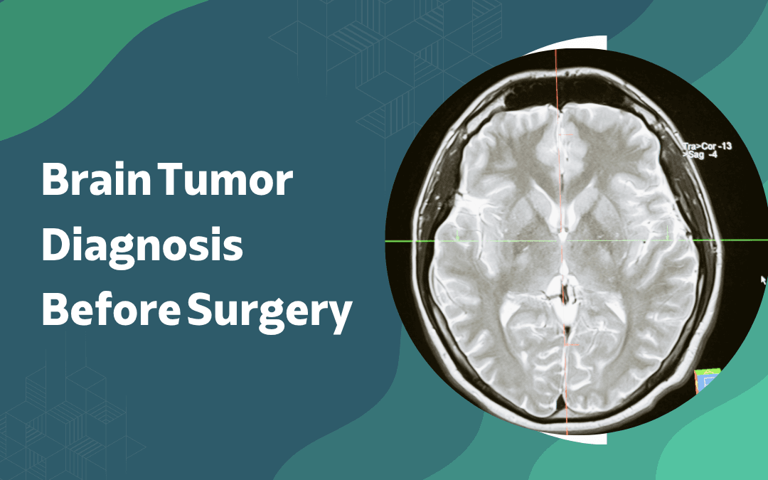
Brain tumor diagnosis before surgery follows a defined sequence: neurological examination, contrast MRI, additional imaging where indicated, and tissue biopsy to confirm histology and molecular grade. Imaging identifies location and likely tumor type biopsy provides the cellular data that determines surgical approach and adjuvant therapy. The workup answers two questions before the patient enters the operating room: what is the tumor, and what can safely be done about it.
According to Dr. Gurneet Singh Sawhney, best neurosurgeon in Mumbai, “Surgery planned without functional MRI and tractography in an eloquent-area tumor isn’t complete planning. The images tell you where the tumor is, but the functional maps tell you what you can actually take.”
Diagnosed with a high-grade glioma and uncertain about the next steps?
What Imaging Tests Are Used to Diagnose a Brain Tumor?
Imaging forms the first and most detailed layer of the pre-surgical diagnostic workup.
Contrast MRI: MRI with gadolinium contrast is the primary modality it identifies tumor location, size, borders, and degree of enhancement, with ring-enhancing patterns pointing toward high-grade malignancy and non-enhancing lesions more consistent with low-grade glioma or other pathology.
CT scan: A CT is faster and more accessible than MRI and remains useful for detecting calcification, acute haemorrhage, or bony skull involvement that MRI can underrepresent — it’s often the first scan ordered in emergency presentations before a planned MRI follows.
Functional MRI: fMRI maps language, motor, and sensory cortex relative to the tumor’s position, giving the surgical team a pre-operative picture of which eloquent areas need to be preserved and whether an awake craniotomy approach is warranted.
PET scan: PET with amino acid tracers differentiates active tumor tissue from treatment-related changes or oedema, particularly useful in recurrent cases where conventional MRI can’t reliably distinguish progression from post-treatment necrosis.
Imaging alone doesn’t confirm tumor type or grade it narrows the differential and informs surgical planning, but tissue diagnosis remains the definitive step. When surgical access to a deep or eloquent lesion is planned, the same imaging stack feeds directly into brain surgery navigation systems used intraoperatively.
What Neurological and Pathological Tests Complete the Pre-Surgical Workup?
Clinical assessment and tissue analysis sit alongside imaging and determine what the surgical team can safely plan.
Neurological exam: Assesses motor strength, reflexes, speech, vision, coordination, and cognition establishing which deficits exist before surgery and a reference point for post-operative function.
Neuropsychological testing: Documents existing cognitive function before surgery near language or memory areas, guides intraoperative mapping, and supports outcome documentation that imaging alone can’t provide.
Stereotactic biopsy: For deep or eloquent lesions where upfront resection isn’t safe, a needle biopsy under CT or MRI guidance confirms histology before any approach to adjacent critical structures feeding functional neurosurgery planning.
Molecular profiling: IDH mutation, MGMT methylation, and 1p/19q co-deletion determine chemotherapy eligibility, radiation dosing, and prognosis in ways histological grade alone cannot.
The pre-surgical workup isn’t finished when imaging is done — it’s finished when histology and molecular data are in hand and the team has a complete picture of both the tumor and the brain around it. For a closer look at how tissue sampling works when imaging is inconclusive, see stereotactic brain biopsy.
Why Choose Dr. Gurneet Singh Sawhney?
Dr. Gurneet Singh Sawhney holds MBBS, MS (General Surgery), and MCh (Neurosurgery) ranking first in the MCh university examination with fellowship training in Functional Neurosurgery and Epilepsy Surgery from Japan, and over 18 years of surgical experience managing brain tumor cases requiring functional MRI, tractography, intraoperative mapping, and stereotactic biopsy as standard pre-surgical workup.
Every brain tumor case is reviewed with imaging before the consultation the surgical discussion is built on the patient’s specific anatomy, not a generic protocol.
Frequently Asked Questions
Is MRI alone enough to diagnose a brain tumor before surgery?
No, MRI identifies the lesion but tissue biopsy confirms tumor type and grade.
What is functional MRI and why is it done before brain tumor surgery?
It maps language and motor areas relative to the tumor to guide safe surgical planning.
Does every brain tumor require a biopsy before surgery?
Not always some tumors are resected directly, with biopsy performed on the removed specimen.
How long does the pre-surgical diagnostic workup typically take?
It varies, but imaging, neurological assessment, and biopsy together usually take one to two weeks.
Disclaimer: The information shared in this content is for educational purposes only and not for promotional use.